EEG re-reference with Brainstorm
This tutorial explains how to re-reference electrode channels (EEG, EOG, ECG) using Brainstorm. We will focus on the case in which EOG or ECG was recorded using unipolar electrodes instead of bipolar electrodes.
Background
EEG is used to measure changes in voltage, which is the difference in electric potential energy between two points in space. For this reason, a single channel of EEG requires two electrodes. The recorded signal is the difference in electric potential between the two electrodes.
In modern EEG, two types of electrode recording configurations are commonly used: unipolar and bipolar.
- In a bipolar configuration, two electrodes are placed on the head, and the resulting signal is the difference between those two electrodes.
- In a unipolar configuration, multiple (more than 3) electrodes are placed on the head, and one of those electrodes serves as a reference for all the others. The recorded signals are then the difference between the remaining electrodes and the reference electrode.
EOG and ECG are commonly recorded using bipolar electrodes, and scalp EEG is commonly recorded using unipolar electrodes.
It is important to note that configuration of how the channels are referenced (commonly called a montage) can be easily changed by subtracting pairs of electrodes from each other. For example, let's say that three electrodes are used at positions F3, F4, M1. In the recording, M1 is used as the reference. This means that the recordings at electrodes F3 and F4 will be the difference in potential between each electrode and M1.
- Channel 1: F3-M1
- Channel 2: F4-M1
If we want to change the reference to F4, then we can subtract the second channel from the first and multiply the second channel by -1.
- New Channel 1: (F3-M1) - (F4-M1)
- New Channel 2: -1*(F4-M1)
This reduces to:
- New Channel 1: F3-F4
- New Channel 2: M1-F4
Unipolar electrodes can also be changed to bipolar by simply subtracting the two electrodes from each other. However, bipolar electrodes cannot commonly be changed to unipolar.
Re-referencing using Brainstorm
Brainstorm allows you to change to a new montage either in the display, during artifact detection, or permanently. An in-depth description of this can be found on the Montage Editor Page.
In the following tutorial, I will focus on the specific case in which the EOG and ECG electrodes were recorded using the unipolar electrode slots instead of the bipolar electrode slots. This requires first changing the montage in the display to check whether everything looks okay and second changing the montage in the artifact detection window. Note that Brainstorm also lets you permanently save the new channels, but this is probably not necessary unless the EOG and ECG are being used for something besides artifact detection.
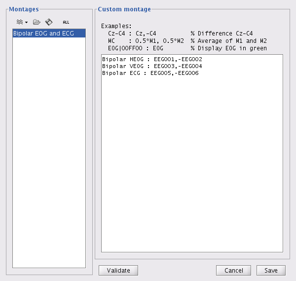
I will assume that HEOG are recorded on channels EEG001 and EEG002, VEOG are recorded on channels EEG003 and EEG004, and ECG are recorded on EEG005 and EEG006. Substitute different channel numbers if your configuration was different.
Changing montage in the display
- After reading in your raw data file, right click on Link to raw file and choose EEG -> Display time series.
- Under the Record tab choose All -> 'Edit montages'. Or right click in the EEG display window and choose Montage -> Edit montages. This brings up the Montage editor.
- Click on the wavy lines under Montages and choose New custom montage. Provide a name for the new montage (e.g., Bipolar EOG and ECG).
- Enter your new montage in the Custom montage box.
- Choose Save.
- The new montage should be displayed in the window and available under the Montage selections.
Changing montage during artifact detection
The montage syntax can be used when selecting the channel to use during artifact detection. For example...
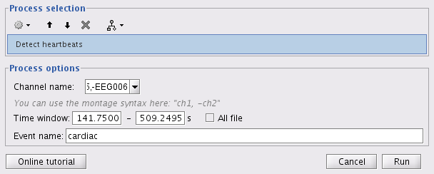
- Choose Artifacts -> Detect heartbeats.
- Under Channel name type in: EEG005,-EEG006
- Brainstorm will use this montage when detecting the cardiac artifacts.

