Lesion-Deficit Correlation Mapping in Aphasia
Funding source: Advancing a Healthier Wisconsin Project 5520462 (Fitzsimmons, PI)
Aphasia is an unfortunately common and often devastating consequence of stroke and other brain injuries. The overall goal of this project is to document relationships between locations of brain damage and specific language processing deficits in people with aphasia. Our hope is that this work will lead to improved classification and diagnosis of aphasia subtypes, methods for early prognostication based on lesion analysis, and improved methods for aphasia rehabilitation. Lesion-deficit correlation also provides information about the brain structures that are critically necessary for a particular cognitive process, which can’t be determined from activation measurements alone. The issue of which brain regions are critically necessary for normal language functions is a central concern in many neurosurgical settings, and large gaps remain in our understanding of these relationships.
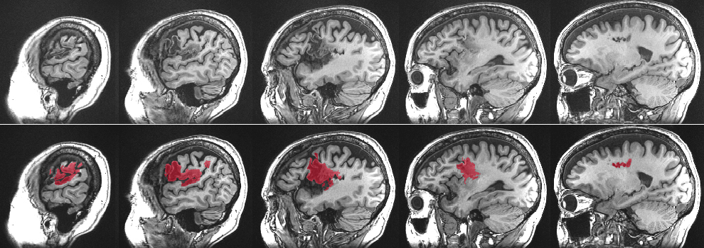
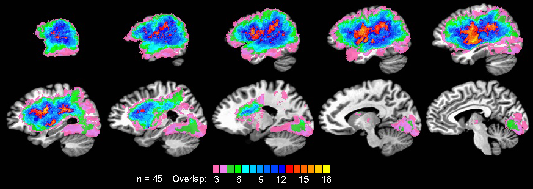
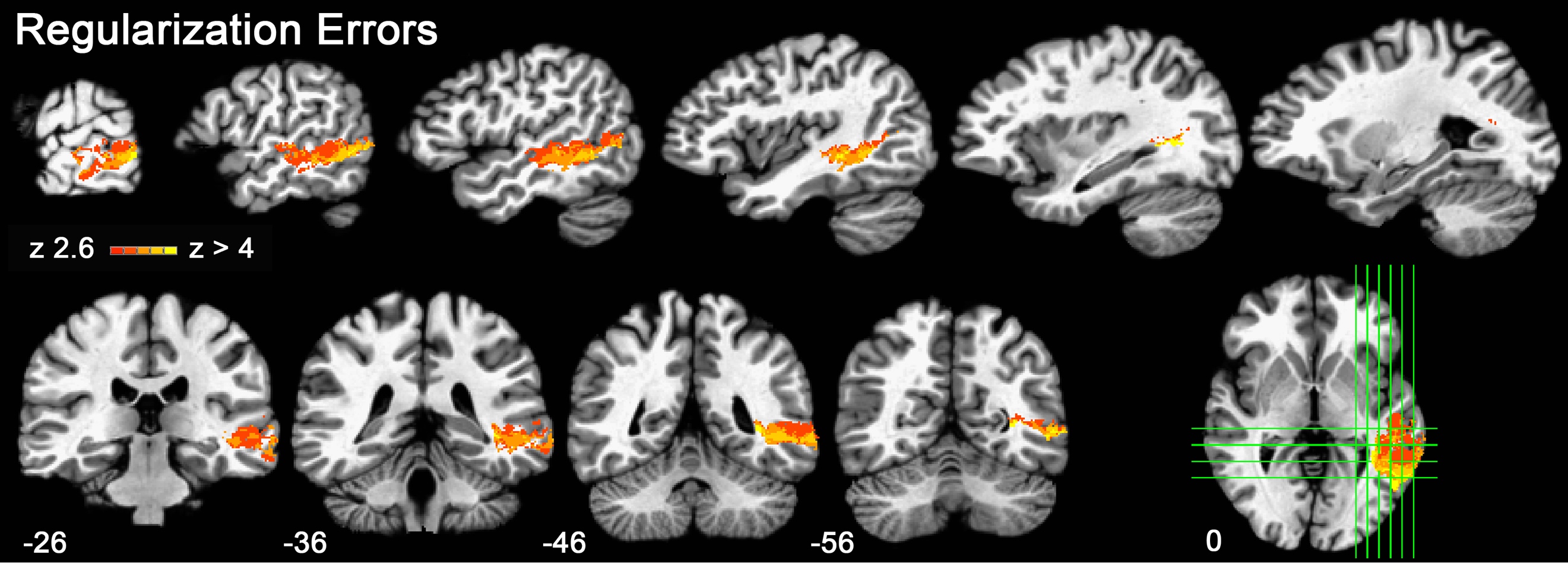
Our work to date has used voxel-based lesion symptom mapping (VLSM), an approach in which the relationship between a specific language deficit and damage at a given voxel is established by dividing a patient sample into those with damage at the voxel and those without damage at the voxel. A statistical comparison (e.g., a t-test) between the scores of the two groups yields a statistical parameter for the voxel, and repetition of this procedure at each voxel produces a statistical parametric map. A defining feature of our VLSM work is the incorporation of behavioral covariates to control for performance variation not specifically related to the language process of interest, such as sensory (auditory or visual), attention, and working memory abilities. In addition to focusing the analysis on specific processes of interest, these controls also help reduce nonspecific effects of variation in lesion size and spatial covariance across voxels due to underlying patterns of the cerebral arterial supply.
Participants undergo an extensive battery of experimental psycholinguistic tests that provide the dependent measures for VLSM. Most of these tests were developed in-house, often as rigorously controlled materials for fMRI experiments in healthy participants. A wide range of language behaviors are assessed, including phoneme and grapheme perception, phonological access from various visual and auditory inputs, phonological short-term memory, phoneme production, object recognition, word comprehension, phrase and sentence comprehension, sentence production, and written word production. A range of experimental manipulations are conducted using these tasks to assess effects of phonological and orthographic structure, lexicality, frequency, orthographic transparency, imageability, semantic category, syntactic structure, and other variables. All tests are administered using computer-controlled stimulus presentations with automated recording of manual choice responses (via a touch-sensitive screen) and vocal responses.
Relevant Publications
- Binder JR, Pillay SB, Humphries CJ, Gross WL, Graves WW, Book DS. Surface errors without semantic impairment in acquired dyslexia: A voxel-based lesion-symptom mapping study. Brain, 2016; 139: 1517-1526.
- Pillay SB, Stengel BC, Humphries C, Book DS, Binder JR. Cerebral localization of impaired phonological retrieval during a rhyme judgment task. Annals of Neurology, 2014; 76: 738-746 (PMCID: PMC4214892).
- Pillay SB, Binder JR, Humphries C, Gross WL, Book DS. Lesion localization of speech comprehension deficits in chronic aphasia. Neurology, 2017; 88: 970-975.